Discovering the Invisible: X-Rays in the 19th Century
The evolution of X-Ray technology has significantly transformed medical diagnostics. From its early adoption, X-Rays provided clear images that dramatically improved the accuracy of diagnoses. Innovations like digital imaging, AI integration, and portable machines offer enhanced efficiency and accessibility in healthcare. These advancements will continue to shape pathology practices, leading to better patient outcomes and personalized treatments.
Did you ever wonder how X-Rays changed the way we see inside our bodies? This incredible discovery revolutionized medicine and made diagnostics more precise and effective…
The discovery of X-Rays
The discovery of X-Rays changed everything in medicine. Wilhelm Röntgen stumbled upon them in 1895. He found that these rays could pass through soft tissues but not bones. This meant doctors could see inside the body without surgery!
This was a breakthrough for diagnosing injuries and diseases. Before X-Rays, doctors had to rely only on physical exams and guesswork. Now, they had a powerful tool to spot fractures, tumors, and other issues.
The First X-Ray
The first X-Ray image Röntgen took was of his wife’s hand. You could see her wedding ring and bones clearly! This simple image amazed everyone and sparked interest all over the world.
How X-Rays Work
X-Rays work by using a special machine that generates these invisible rays. When the rays pass through the body, they create images on a film or digital sensor. Dense materials like bones appear white, while softer areas look darker.
Impact on Medicine
The impact on medicine has been huge. X-Rays allow doctors to see what’s happening inside the body without being invasive. It has led to quicker diagnoses and better treatments. X-Rays are now commonplace in hospitals and clinics.
Thanks to the discovery of X-Rays, we have come to trust imaging technologies to guide our healthcare decisions. This advancement paved the way for even more diagnostic tools, like MRIs and CT scans.
Impact on medical diagnostics
The impact of X-Rays on medical diagnostics has been profound. Before their invention, doctors often guessed what might be wrong with patients. This led to mistakes and delays in treatment. With X-Rays, a clear view of the inside of the body became possible.
Now, doctors can spot broken bones, tumors, and foreign objects quickly. This technology helps in guiding surgeries and other treatments. Patients no longer need to undergo invasive procedures just to see inside their bodies.
Improved Accuracy
X-Rays have greatly improved diagnostic accuracy. Doctors can make better decisions based on clear images. They rely on these images to understand complex health issues. This not only saves time but also increases patient trust.
Health Screenings
X-Rays are also used in routine health screenings. For instance, chest X-Rays can detect lung diseases early. Regular screenings are vital for preventing serious health conditions.
Advantages Over Traditional Methods
Compared to older methods, X-Rays are faster and safer. Traditional methods required exploratory surgeries, which were risky. X-Rays allow doctors to see what’s wrong with much less risk.
The ongoing advancements in X-Ray technology continue to shape the future of diagnostics. They allow for innovations like digital X-Rays, which use less radiation and provide immediate results. This means quicker diagnosis and treatment for patients.
Evolution of pathology practices
The evolution of pathology practices thanks to X-Rays has been remarkable. In the past, doctors had to rely on visible symptoms and basic tests to diagnose illnesses. This often led to delays in treatment and misdiagnoses.
With the introduction of X-Rays, pathologists began to access a new world of information. They could visualize internal structures and identify problems that were previously hidden. This ability to see inside the body significantly improved diagnosis accuracy.
New Techniques
As X-Ray technology advanced, so did the techniques used in pathology. Medical professionals started using X-Rays to study tissue samples and to guide biopsies. This allowed for faster and more precise diagnoses.
Integration with Other Technologies
Pathology practices have also integrated X-Rays with other imaging technologies. Methods like CT scans and MRIs complement X-Rays. Together, these technologies provide a complete picture for diagnosing diseases.
Impact on Treatment Options
The ability to diagnose conditions accurately led to better treatment options. Doctors now tailor treatments based on what they see in the X-Ray images. This personalization boosts recovery rates and enhances patient care.
Overall, X-Rays have transformed pathology from basic approaches to a sophisticated science. Now, pathologists play a crucial role in patient diagnosis and treatment. Their expertise, combined with advanced imaging, drives healthcare forward.
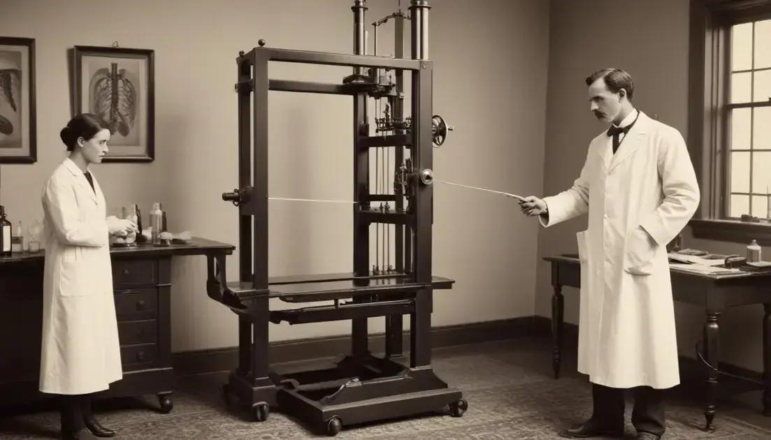
Challenges in early adoption of X-Rays
Early adoption of X-Rays wasn’t easy. Many doctors were unsure about this new technology. They worried about the safety of using X-Rays. The idea of radiation exposure was frightening for many healthcare workers and patients alike.
Another challenge was the lack of training. Most doctors had no experience with X-Ray machines. This made it hard for them to use the technology correctly. Mistakes could lead to inaccurate diagnoses.
Cost of Equipment
The cost of X-Ray machines was also a barrier. Many small clinics couldn’t afford this expensive technology. This kept X-Rays from being widely available in the beginning.
Public Perception
Public perception played a role too. Some patients were afraid of the unknown. They didn’t trust a machine to see inside their bodies. This fear slowed down the acceptance of X-Ray technology.
Over time, education and successful case studies helped overcome these challenges. As more doctors saw the benefits, they became more willing to adopt X-Rays. The advancements in X-Ray technology have proven essential for modern medicine.
Future of X-Ray technology
The future of X-Ray technology looks bright and exciting. New advances are making X-Rays safer and more effective. For example, digital X-Rays are becoming more common. They use less radiation and provide instant images for doctors.
Another trend is the development of portable X-Ray machines. These devices allow healthcare professionals to take X-Rays anywhere, even in remote areas. This means more people can access essential diagnostic tools.
AI Integration
Artificial intelligence (AI) is also playing a big role. AI can help doctors analyze X-Ray images faster and more accurately. This technology can spot problems that even trained eyes might miss.
3D Imaging
3D imaging is another exciting advancement. This technology enables doctors to see detailed, three-dimensional views of bones and tissues. It helps in planning surgeries with greater precision.
Overall, the future developments in X-Ray technology promise to improve patient care. With ongoing research and innovation, healthcare will become more efficient and accessible. X-Rays will continue to be a vital tool in diagnostics and treatment.
Conclusion
In conclusion, the evolution of X-Ray technology has significantly impacted medicine and diagnostics. From the early days of its discovery to the advanced techniques we see today, X-Rays have changed how doctors diagnose and treat patients. They provide quick and accurate images that help in making informed decisions.
As we look to the future, innovations like digital imaging, portable machines, and AI integration promise to make X-Rays even more effective and accessible. These advancements will continue to improve patient care and make healthcare more efficient.
Ultimately, understanding the journey of X-Ray technology helps us appreciate its role in modern medicine. As technology progresses, so will the potential of X-Rays to enhance diagnostics and treatments for patients everywhere.